Skip to content Skip to sidebar Skip to footer

Wilson Disease Mri Brain

Wilson Disease Radiology Reference Article Radiopaedia Org

Wilson Disease Radiology Reference Article Radiopaedia Org

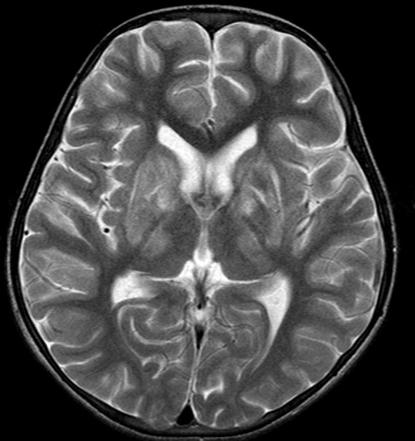
Wilson disease mri brain. MRI studies have identified focal abnormalities in the white matter pons and deep. Positive findings believed secondary to this condition were found in 15 subjects. Wilson disease hepatolenticular degeneration is an autosomal recessive defect in cellular copper transport.

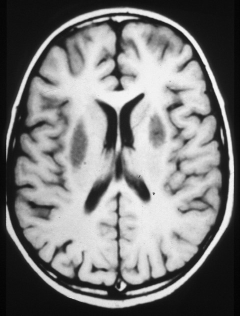
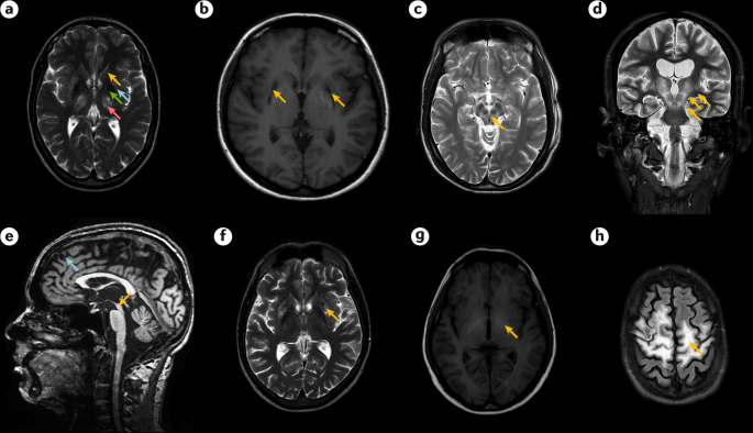
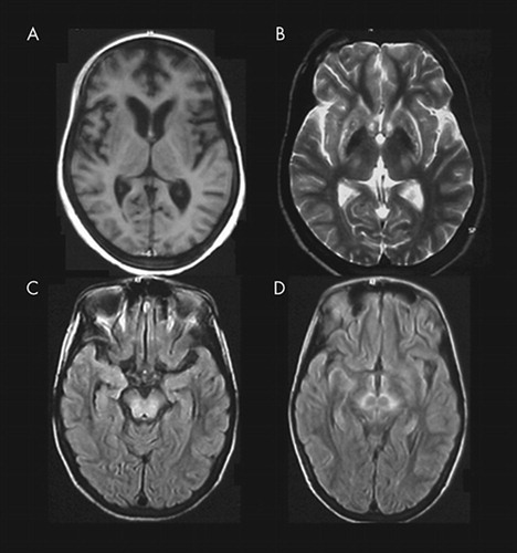
6 8 Neurologic WD is one of the main forms of the disease. In Wilson disease WD T2T2weighted T2w MRI frequently shows hypointensity in the basal ganglia that is suggestive of paramagnetic deposits. It is currently unknown whether this hypointensity is related to copper or iron deposition.

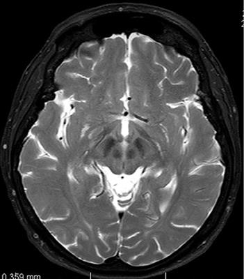
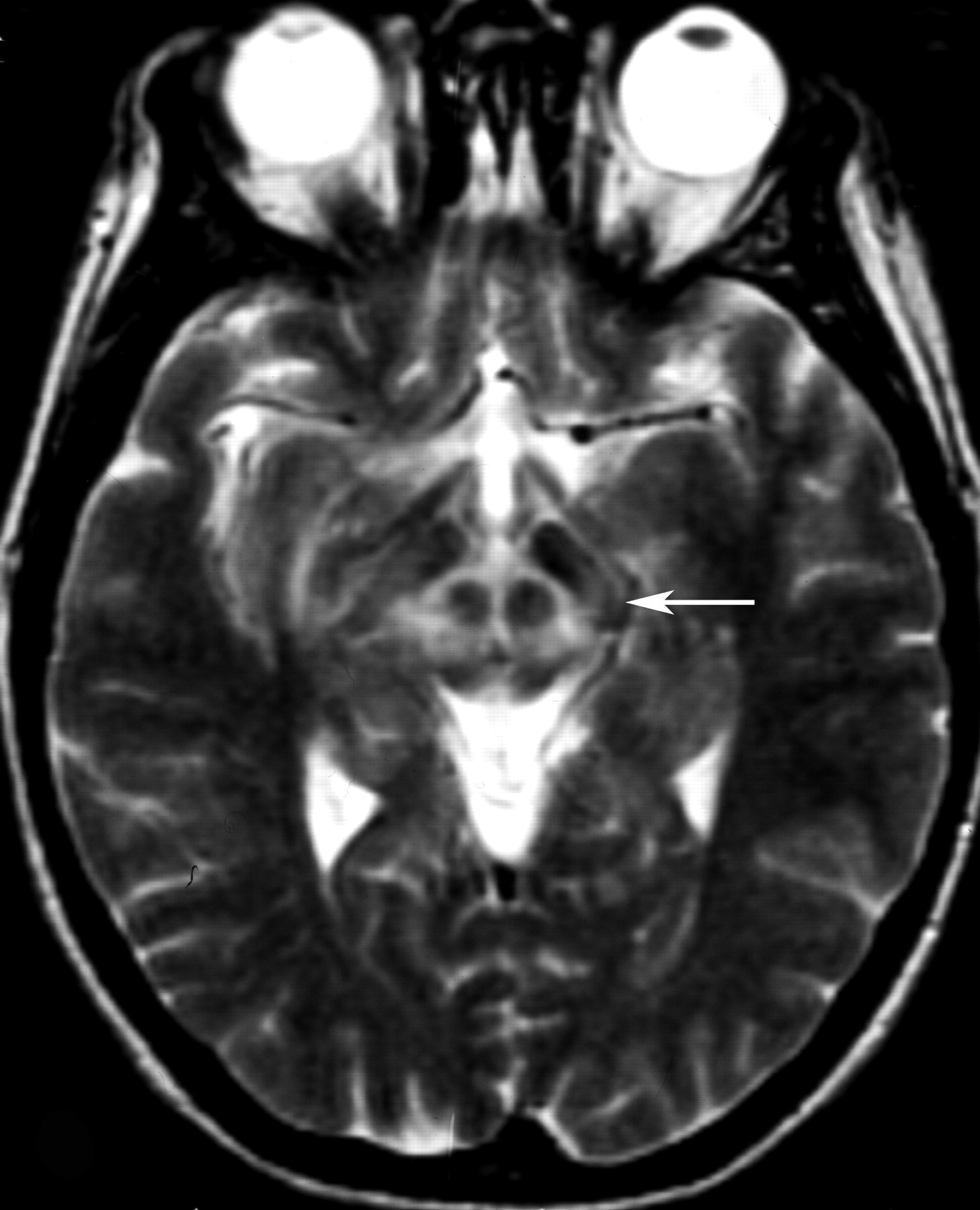
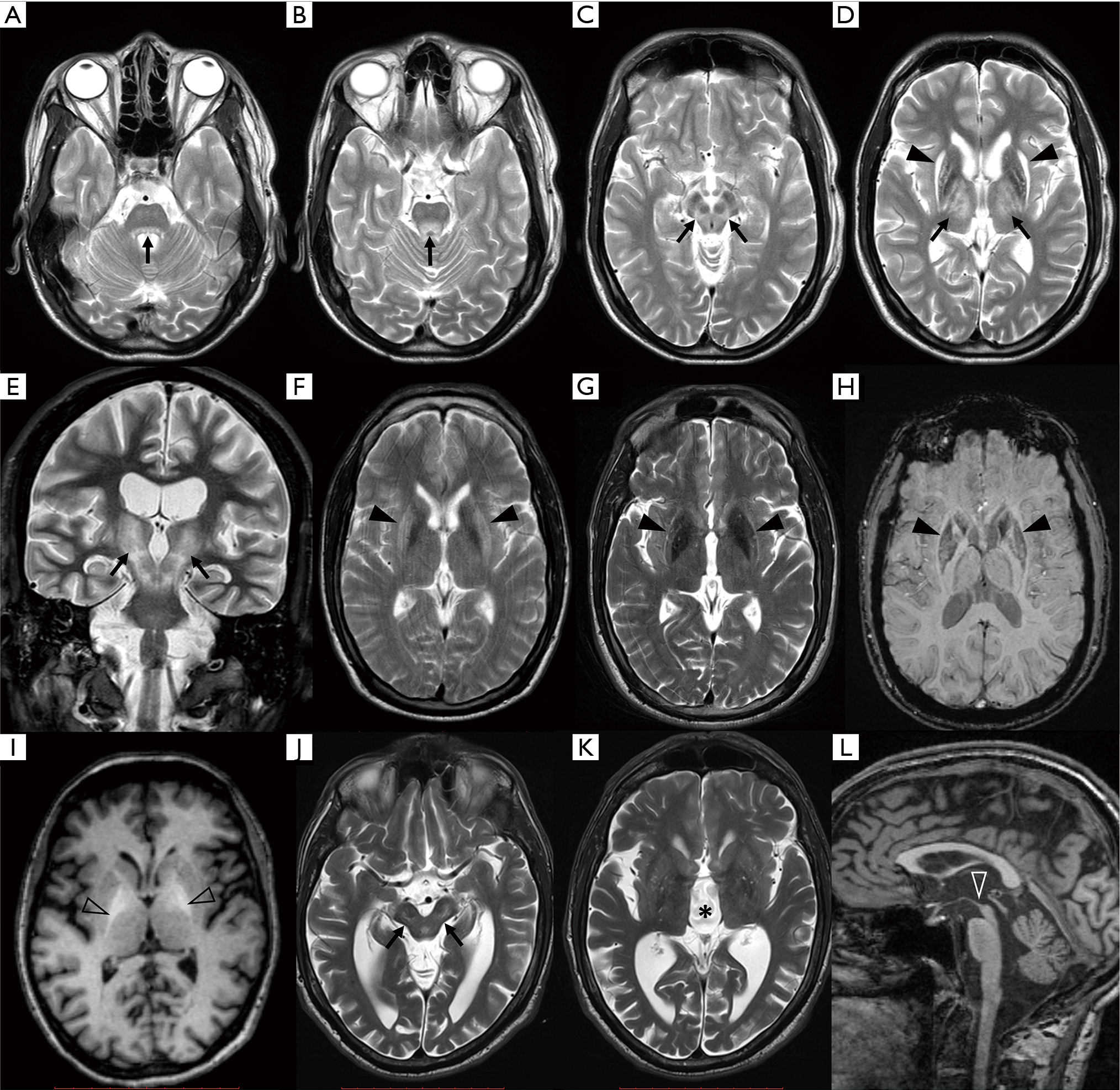
Interval changes on follow-up MR imaging were also closely correlated with clinical findings and. MRI of the brain appears to be more sensitive than CT scanning in detecting early lesions of Wilson disease. The face of the giant panda sign panda sign of the midbrain or double-panda sign is a characteristic pandas face appearance in magnetic resonance imaging MRI images of people with Wilsons disease.

This article aims to discuss the central nervous system manifestations of this condition. It is found worldwide with a prevalence of approximately 1 case in 30000 live births in most populations. Dysarthria tremor ataxia rigiditybradykinesia and choreadystonia.

Wilsons disease is an inherited disorder in which defective biliary excretion of copper leads to its accumulation particularly in the liver and brain. The patients were scanned using spin-echo SE sequences. Wilsons disease WD is due to excessive copper accumulation in the liver and brain 1.

The neurologist was looking for typical symptoms. Wilson disease also known as hepatolenticular degeneration is a multisystem disease due to abnormal accumulation of copper. It is characterized by early onset liver cirrhosis with CNS findings most frequently affecting the basal ganglia and midbrain.

1 In addition a second miniature panda face can be seen in the high signal abnormality in the pons figure C. To describe the spectrum of brain abnormalities in Wilson disease hepatolenticular degeneration as depicted at magnetic resonance MR imaging and computed tomography CT and to relate these findings to neurologic and hepatologic abnormalities.

Mr Imaging Of The Brain In Wilson Disease Of Childhood Findings Before And After Treatment With Clinical Correlation American Journal Of Neuroradiology

Mr Imaging Of The Brain In Wilson Disease Of Childhood Findings Before And After Treatment With Clinical Correlation American Journal Of Neuroradiology

Mr Imaging Of The Brain In Wilson Disease Of Childhood Findings Before And After Treatment With Clinical Correlation American Journal Of Neuroradiology

Mr Imaging Of The Brain In Wilson Disease Of Childhood Findings Before And After Treatment With Clinical Correlation American Journal Of Neuroradiology

Atypical Brain Mri Findings In Wilson S Disease Patients Extensive Download Scientific Diagram

Atypical Brain Mri Findings In Wilson S Disease Patients Extensive Download Scientific Diagram

Wilson Disease Radiology Reference Article Radiopaedia Org

Wilson Disease Radiology Reference Article Radiopaedia Org

Scielo Brasil Frontal Lobes White Matter Abnormalities Mimicking Cystic Leukodystrophy In Wilson S Disease Frontal Lobes White Matter Abnormalities Mimicking Cystic Leukodystrophy In Wilson S Disease

Scielo Brasil Frontal Lobes White Matter Abnormalities Mimicking Cystic Leukodystrophy In Wilson S Disease Frontal Lobes White Matter Abnormalities Mimicking Cystic Leukodystrophy In Wilson S Disease

Frontiers Late Onset Wilson S Disease Medicine

Frontiers Late Onset Wilson S Disease Medicine

Mr Imaging Of The Brain In Wilson Disease Of Childhood Findings Before And After Treatment With Clinical Correlation American Journal Of Neuroradiology

Mr Imaging Of The Brain In Wilson Disease Of Childhood Findings Before And After Treatment With Clinical Correlation American Journal Of Neuroradiology

Classical Brain Mri Findings In Wilson S Disease Patients Bilateral Download Scientific Diagram

Classical Brain Mri Findings In Wilson S Disease Patients Bilateral Download Scientific Diagram

Presentation1 Radiological Imaging Of Wilson Disease

Presentation1 Radiological Imaging Of Wilson Disease

Wilson Disease Radiology Reference Article Radiopaedia Org

Wilson Disease Radiology Reference Article Radiopaedia Org

Atypical Brain Mri Findings In Wilson S Disease Patients Extensive Download Scientific Diagram

Atypical Brain Mri Findings In Wilson S Disease Patients Extensive Download Scientific Diagram

A Study Of Mri Changes In Wilson Disease And Its Correlation With Clinical Features And Outcome Sciencedirect

A Study Of Mri Changes In Wilson Disease And Its Correlation With Clinical Features And Outcome Sciencedirect

Mr Imaging Of The Brain In Wilson Disease Of Childhood Findings Before And After Treatment With Clinical Correlation American Journal Of Neuroradiology

Mr Imaging Of The Brain In Wilson Disease Of Childhood Findings Before And After Treatment With Clinical Correlation American Journal Of Neuroradiology

Wilson Disease Radiology Case Radiopaedia Org

Wilson Disease Radiology Case Radiopaedia Org

A Study Of Brain Mri Characteristics And Clinical Features In 76 Cases Of Wilson S Disease Sciencedirect

A Study Of Brain Mri Characteristics And Clinical Features In 76 Cases Of Wilson S Disease Sciencedirect

Wilson S Disease And Other Neurological Copper Disorders The Lancet Neurology

Wilson S Disease And Other Neurological Copper Disorders The Lancet Neurology

Cranial Mri Of A Wilson Disease Patient Presenting With Cognitive Download Scientific Diagram

Cranial Mri Of A Wilson Disease Patient Presenting With Cognitive Download Scientific Diagram

The Double Panda Sign And Central Pontine Signal Changes In Wilson S Disease Eurorad

The Double Panda Sign And Central Pontine Signal Changes In Wilson S Disease Eurorad

Brain Magnetic Resonance Spectroscopy Mrs As A Diagnostic Tool For Detecting Early Neurological Changes In Children With Wilson S Disease European Journal Of Radiology

Brain Magnetic Resonance Spectroscopy Mrs As A Diagnostic Tool For Detecting Early Neurological Changes In Children With Wilson S Disease European Journal Of Radiology

Presentation1 Radiological Imaging Of Wilson Disease

Presentation1 Radiological Imaging Of Wilson Disease

Classic Neuroimaging The Bird S Eye View In Wilson S Disease Bmj Case Reports

Classic Neuroimaging The Bird S Eye View In Wilson S Disease Bmj Case Reports

Epos C 1459

Epos C 1459

Wilson Disease Cns Manifestations Radiology Reference Article Radiopaedia Org

Wilson Disease Cns Manifestations Radiology Reference Article Radiopaedia Org

1

1

Presentation1 Radiological Imaging Of Wilson Disease

Presentation1 Radiological Imaging Of Wilson Disease

Atypical Mri Features Involving The Brain In Wilson S Disease Abstract Europe Pmc

Atypical Mri Features Involving The Brain In Wilson S Disease Abstract Europe Pmc

Fig 5 Mr Imaging Of The Brain In Wilson Disease Of Childhood Findings Before And After Treatment With Clinical Correlation American Journal Of Neuroradiology

Fig 5 Mr Imaging Of The Brain In Wilson Disease Of Childhood Findings Before And After Treatment With Clinical Correlation American Journal Of Neuroradiology

Http Pdf Posterng Netkey At Download Index Php Module Get Pdf By Id Poster Id 105811

Http Pdf Posterng Netkey At Download Index Php Module Get Pdf By Id Poster Id 105811

Brain Mri And Spect In The Diagnosis Of Early Neurological Involvement In Wilson S Disease Semantic Scholar

Brain Mri And Spect In The Diagnosis Of Early Neurological Involvement In Wilson S Disease Semantic Scholar

Pdf Wilson S Disease A 2017 Update

Pdf Wilson S Disease A 2017 Update

Neurologic Impairment In Wilson Disease Dusek Annals Of Translational Medicine

Neurologic Impairment In Wilson Disease Dusek Annals Of Translational Medicine

Http Pdf Posterng Netkey At Download Index Php Module Get Pdf By Id Poster Id 105811

Http Pdf Posterng Netkey At Download Index Php Module Get Pdf By Id Poster Id 105811

Figure 2 From Atypical Mri Features Of Wilson S Disease High Signal In Globus Pallidus On T1 Weighted Images Semantic Scholar

Figure 2 From Atypical Mri Features Of Wilson S Disease High Signal In Globus Pallidus On T1 Weighted Images Semantic Scholar

Teaching Neuroimages Mri Reversal In Wilson Disease With Trientine Treatment Neurology

Teaching Neuroimages Mri Reversal In Wilson Disease With Trientine Treatment Neurology

Kathy S Mri Pathology Blog Wilson S Disease

Kathy S Mri Pathology Blog Wilson S Disease

Trident And Face Of Giant Panda Sign On Mri In Wilsons Disease Note Deposition Of Copper In Basal Ganglia

Trident And Face Of Giant Panda Sign On Mri In Wilsons Disease Note Deposition Of Copper In Basal Ganglia

Wilson Disease Nature Reviews Disease Primers

Wilson Disease Nature Reviews Disease Primers

Wilson S Disease Mri Features Abstract Europe Pmc

Wilson S Disease Mri Features Abstract Europe Pmc

Figure 1 From Atypical Mri Features Of Wilson S Disease High Signal In Globus Pallidus On T1 Weighted Images Semantic Scholar

Figure 1 From Atypical Mri Features Of Wilson S Disease High Signal In Globus Pallidus On T1 Weighted Images Semantic Scholar

Classic Neuroimaging The Bird S Eye View In Wilson S Disease Bmj Case Reports

Classic Neuroimaging The Bird S Eye View In Wilson S Disease Bmj Case Reports

Face Of The Giant Panda Sign Wikipedia

Face Of The Giant Panda Sign Wikipedia

Dr Balaji Anvekar Frcr Wilson Disease Mri

Dr Balaji Anvekar Frcr Wilson Disease Mri

Wilson Disease Chapter 5 Uncommon Causes Of Movement Disorders

Wilson Disease Chapter 5 Uncommon Causes Of Movement Disorders

Parkinsonism Related Disorders

Parkinsonism Related Disorders

Psychiatric Manifestations In Wilson S Disease A Cross Sectional Analysis The Journal Of Neuropsychiatry And Clinical Neurosciences

Psychiatric Manifestations In Wilson S Disease A Cross Sectional Analysis The Journal Of Neuropsychiatry And Clinical Neurosciences

A Study Of Brain Mri Characteristics And Clinical Features In 76 Cases Of Wilson S Disease Sciencedirect

A Study Of Brain Mri Characteristics And Clinical Features In 76 Cases Of Wilson S Disease Sciencedirect

Wilson S Disease Diagnosis Of Wilson S Disease In Ethiopian Young Sisters

Wilson S Disease Diagnosis Of Wilson S Disease In Ethiopian Young Sisters

Http Pdf Posterng Netkey At Download Index Php Module Get Pdf By Id Poster Id 105811

Http Pdf Posterng Netkey At Download Index Php Module Get Pdf By Id Poster Id 105811

1

1

The neurologist was looking for typical symptoms.

The National Organizations for Rare Disorders NORD reported that 1 in every 30000 to 40000 people in the world are affected by WD 1. To describe the spectrum of brain abnormalities in Wilson disease hepatolenticular degeneration as depicted at magnetic resonance MR imaging and computed tomography CT and to relate these findings to neurologic and hepatologic abnormalities. 3 5 WD leads to intracellular copper accumulation causing damage to many organs especially the brain. Dysarthria tremor ataxia rigiditybradykinesia and choreadystonia. In Wilson disease WD T2T2weighted T2w MRI frequently shows hypointensity in the basal ganglia that is suggestive of paramagnetic deposits. Wilson disease also known as hepatolenticular degeneration is a multisystem disease due to abnormal accumulation of copper. It is found worldwide with a prevalence of approximately 1 case in 30000 live births in most populations. Patients neurological symptoms brain MRI features and laboratory findings were noted. The patients were scanned using spin-echo SE sequences.


To describe the spectrum of brain abnormalities in Wilson disease hepatolenticular degeneration as depicted at magnetic resonance MR imaging and computed tomography CT and to relate these findings to neurologic and hepatologic abnormalities. Positive findings believed secondary to this condition were found in 15 subjects. It is found worldwide with a prevalence of approximately 1 case in 30000 live births in most populations. Thirty-eight patients with biochemically proven Wilsons disease underwent magnetic resonanceimaging MRI of the brain as well as neurological examinations. Fifty patients with Wilson disease participated in the cross-sectional. The face of the giant panda sign panda sign of the midbrain or double-panda sign is a characteristic pandas face appearance in magnetic resonance imaging MRI images of people with Wilsons disease. 1 In addition a second miniature panda face can be seen in the high signal abnormality in the pons figure C.

Post a Comment for "Wilson Disease Mri Brain"